![]() BG - Brazilian Group
BG - Brazilian Group
Nosocomial infections are those that originate in hospitals. In US, such infections affect about 10% of patients and cause more than 20,000 deaths per year. In Brazil, about 45,000 deaths are recorded annually resulting from infections acquired in hospitals in a total of 12 million hospitalizations. The occurrence of these infections is attributed to the high prevalence of
pathogens in predisposed patients (such as immunosupressed or those with lesions in the skin or mucosa) and efficient transmission mechanisms from one patient to another. Nosocomial infections are caused generally by opportunistic microorganisms with prevalence of Grampositive MRSA ( "methicillin-resistant Staphylococcus aureus"), VISA / VRSA ("vancomycin-intermediate / resistant Staphylococcus aureus") and VRE ( "vancomycinresistant enterococci) and Gram-negative bacteria Pseudomonas aeruginosa, Acinetobacter spp., Enterobacteriaceae and producers of extended spectrum beta-lactamases Stenotrophomonas (ESBL). [Deege, M.P.D., and Paterson, D.L., Reducing the Development of Antibiotic Resistance in Critical Care Units. Current Pharmaceutical Biotechnology, 2011.12(12): p. 2062-2069.]
Many species of bacteria can live in free (planktonic) form or parasitizing a host; they should be able to adapt to sudden changes in nutrient availability and host defense systems. A mechanism for the adaptation of bacteria to that effect is its ability to grow as part of a biofilm - a community encapsulated by exo-polymers. Scientific interest in the process of biofilm
formation has escalated in recent years; molecular genetic studies of biofilm formation have begun to indicate the causes for this process. Today it is widely accepted that biofilm formation is an important aspect of many bacterial diseasesi. Biofilms can tolerate antimicrobial agents in concentrations 10-1000 times higher than those needed to kill genetically equivalent planktonic bacteria, thus making them very difficult to be eradicated in live hosts [R.M. Donlan and J.W.Costerton, Biofilms: survival mechanisms of clinically relevant microorganisms. Clin. Microbiol. Rev. 15, 167 (2002)]. Biofilm formation is a process encompassing several different stages starting with the adhesion at the surface, either biotic or abiotic in nature, bacterial growth, and expansion within microcolonies to form highly organized structures.
Thus, in order to eliminate biofilm and related infections, two approaches are necessary. Fast and sensitive detection for early treatment, and a thorough, basic understanding of cell adhesion, cell-cell signaling and biofilm formation.
In this project, the Brazilian team will work to achieve two main objectives: Implementation of high sensitivity biosensors for neglected diseases and those specific for BRICS countries:
We will pursue the fabrication of high sensitivity FET-like biosensors based on the the high surface/volume of nanomaterials; we will use the partners expertise to improve the timeframe of device fabrication (which can take over one year using Brazilian multi-user facilities). Despite the obvious impact on biomedicine, these high sensitivity devices can also be used for in-situ monitoring the production of biomolecules by cells during adhesion and biofilm
formation processes.
Basic studies of cell adhesion, cell-cell signaling and biofilm formation:
Using advanced microscopy, the entire biofilm formation process has been validated, and yet undiscovered stages were found, for the phytopathogen Xylella fastidiosa. With semiconductor nanowire arrays, cell adhesion forces were quantitatively measured and cell movement could be tracked. These studies can provide insights and be extended to human bacteria, using new devices developed in collaboration with the partners in this proposal, such as nano-oscillators and nano-swimmers. In order to fulfill those objectives, the Brazilian team will rely on devices fabricated by their partners and contribute with its expertise on the nanobiointerface. Examples of the methodology used by the Brazilian team are discussed below.
Methods of Biosensor fabrication:
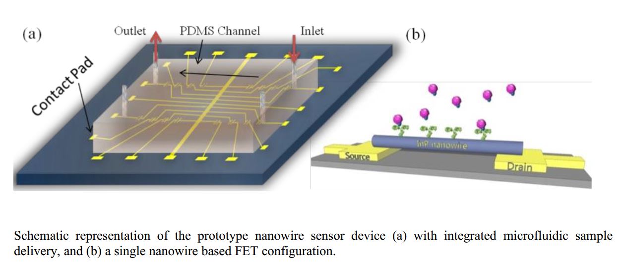
We will develop functionalization protocols and further device fabrication for the different nanomaterials used by our partners for the FET fabrication. For that, prior to target DNA or protein detection, the sample must be processed through a series of functionalization protocols previously used in our group: 1) creating amino groups on surfaces, 2) rational attachment of heterobifunctional PEG linker to the aminated surface, and 3) covalent coupling of biomolecules through peptide binding. A schematic representation of a nanowire sensor device integration with PDMS based microfluidic channels in a chip is depicted in the figure below.
The collaboration with state-run FIOCRUZ (http://portal.fiocruz.br/en/content/home-inglês), the most prominent science and technology health institution in Latin America, will allow access to important proteins of diseases usually neglected by large pharmaceutical companies.
Advanced microscopy for cell adhesion studies:
Ex-vivo studies of cell adhesion are based on the use of advanced microscopy (equipments are listed separately). However, we have configured experiments to quantitatively probe the adhesion process using nano-objects such as nanowire arrays. Each experiment is configured using new methodologies depending on the scientific case to be explored. For this proposal,
for example, nano-oscillators can be used to probe, at very low levels, the local deposition of EPS (extracellular polymeric substances) produced by the cells, by measuring the frequency change due to the extra mass. Nano-swimmers can be used to create a ‘movable’ chemical signal gradient to the planktonic cells if the magnetic particle is properly functionalized.
These experiments can be coupled to microfluidics and observed in real time using fluorescence microscopy.
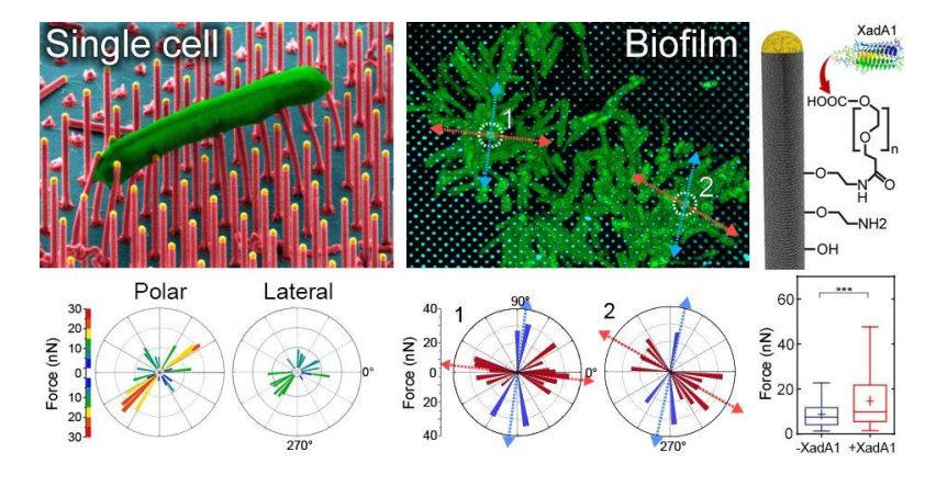
InP nanowire arrays used for bacterial cell adhesion studies. The force value is obtained from the deflection of the nanowire observed by scanning electron microscopy or confocal laser scanning microscopy. Functionalization protocols were developed to probe the role of an adhesin produced by the cells. [P.Sahoo et al., Nano Letters (2016)].